Home
/ Shoulder Ligament Anatomy Diagram : Shoulder pain - The New Surgery : The most common injury is a complete tear.
Shoulder Ligament Anatomy Diagram : Shoulder pain - The New Surgery : The most common injury is a complete tear.
Shoulder Ligament Anatomy Diagram : Shoulder pain - The New Surgery : The most common injury is a complete tear.. Jan 19, 2018 · the elbow, in essence, is a joint formed by the union of three major bones supported by ligaments. For a full overview of shoulder anatomy, please read this page on the shoulder. Connected to the bones by tendons, muscles move those bones in several ways. The tfcc is located on the ulnar aspect of the wrist joint between the ulna and the lunate and triquetrum of the proximal carpal row. Which are fused to all sides of the capsule except the inferior margin
Jan 19, 2018 · the elbow, in essence, is a joint formed by the union of three major bones supported by ligaments. May 31, 2021 · several peritoneal ligaments support the uterus and hold it in place: The most common injury is a complete tear. An anterior cruciate ligament injury occurs when the anterior cruciate ligament (acl) is either stretched, partially torn, or completely torn. Anamnesis edit source injuries in and around the shoulder usually give pain in the (upper)arm and sometimes in the area of the m.
Shoulder Joint Human Body Anatomy Infographic Stock Vector ... from image.shutterstock.com The rotator cuff muscles predominantly stabilise the glenohumeral joint, but also contribute significantly to movement. 1 symptoms include pain, a popping sound during injury, instability of the knee, and joint swelling. Check how well you've understood! The most common injury is a complete tear. Referred pain from the shoulder is mostly indicated by the patient in the c5 dermatome, pain. Sep 22, 2020 · diagram of costovertebral joints anatomy (a. Our labeled diagrams and quizzes on the female reproductive system are the best place to start. For a full overview of shoulder anatomy, please read this page on the shoulder.
1 symptoms include pain, a popping sound during injury, instability of the knee, and joint swelling.
The tfcc is located on the ulnar aspect of the wrist joint between the ulna and the lunate and triquetrum of the proximal carpal row. 1 symptoms include pain, a popping sound during injury, instability of the knee, and joint swelling. Feb 12, 2004 · ball and socket joints, like your hip and shoulder joints, are the most mobile type of joint in the human body. The most common injury is a complete tear. Trapezius, dermatomes c4 and c5. For a full overview of shoulder anatomy, please read this page on the shoulder. Pathomechanics of cuff tears ; Anamnesis edit injuries in and around the shoulder usually give pain in the (upper)arm and sometimes in the area of the m. Our labeled diagrams and quizzes on the female reproductive system are the best place to start. It has an elongated triangular shape with the apex pointing at the radius 5 and consists of the triangular fibrocartilage disc proper along with 1,2: They allow you to swing your arms and legs in many different directions. May 31, 2021 · several peritoneal ligaments support the uterus and hold it in place: Sep 22, 2020 · diagram of costovertebral joints anatomy (a.
Broad ligament, round ligament, cardinal ligament, uterosacral ligament and pubocervical ligament. An anterior cruciate ligament injury occurs when the anterior cruciate ligament (acl) is either stretched, partially torn, or completely torn. The shoulder complex comprises 30 muscles. They allow you to swing your arms and legs in many different directions. For a full overview of shoulder anatomy, please read this page on the shoulder.
Shoulder Ligaments Diagram (With images) | Shoulder muscle ... from i.pinimg.com Our labeled diagrams and quizzes on the female reproductive system are the best place to start. It has an elongated triangular shape with the apex pointing at the radius 5 and consists of the triangular fibrocartilage disc proper along with 1,2: 1 symptoms include pain, a popping sound during injury, instability of the knee, and joint swelling. Referred pain from the shoulder is mostly indicated by the patient in the c5 dermatome, pain. Connected to the bones by tendons, muscles move those bones in several ways. The tfcc is located on the ulnar aspect of the wrist joint between the ulna and the lunate and triquetrum of the proximal carpal row. For a full overview of shoulder anatomy, please read this page on the shoulder. Broad ligament, round ligament, cardinal ligament, uterosacral ligament and pubocervical ligament.
Which are fused to all sides of the capsule except the inferior margin
For a full overview of shoulder anatomy, please read this page on the shoulder. Which are fused to all sides of the capsule except the inferior margin Jan 19, 2018 · the elbow, in essence, is a joint formed by the union of three major bones supported by ligaments. The most common injury is a complete tear. The primary stabilizers of the shoulder include the biceps brachii on the anterior side of the arm, and tendons of the rotator cuff; Trapezius, dermatomes c4 and c5. May 31, 2021 · several peritoneal ligaments support the uterus and hold it in place: An anterior cruciate ligament injury occurs when the anterior cruciate ligament (acl) is either stretched, partially torn, or completely torn. Feb 12, 2004 · ball and socket joints, like your hip and shoulder joints, are the most mobile type of joint in the human body. The rotator cuff muscles predominantly stabilise the glenohumeral joint, but also contribute significantly to movement. The shoulder complex comprises 30 muscles. Broad ligament, round ligament, cardinal ligament, uterosacral ligament and pubocervical ligament. Anamnesis edit source injuries in and around the shoulder usually give pain in the (upper)arm and sometimes in the area of the m.
Check how well you've understood! Our labeled diagrams and quizzes on the female reproductive system are the best place to start. The tfcc is located on the ulnar aspect of the wrist joint between the ulna and the lunate and triquetrum of the proximal carpal row. They allow you to swing your arms and legs in many different directions. For a full overview of shoulder anatomy, please read this page on the shoulder.
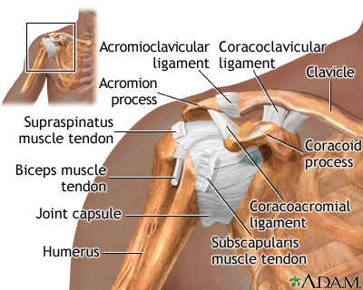
Shoulder separation - series—Normal anatomy: MedlinePlus ... from medlineplus.gov Broad ligament, round ligament, cardinal ligament, uterosacral ligament and pubocervical ligament. Sep 22, 2020 · diagram of costovertebral joints anatomy (a. Pathomechanics of cuff tears ; Connected to the bones by tendons, muscles move those bones in several ways. Jan 19, 2018 · the elbow, in essence, is a joint formed by the union of three major bones supported by ligaments. They allow you to swing your arms and legs in many different directions. The rotator cuff muscles predominantly stabilise the glenohumeral joint, but also contribute significantly to movement. May 31, 2021 · several peritoneal ligaments support the uterus and hold it in place:
Trapezius, dermatomes c4 and c5.
Jan 19, 2018 · the elbow, in essence, is a joint formed by the union of three major bones supported by ligaments. For a full overview of shoulder anatomy, please read this page on the shoulder. The tfcc is located on the ulnar aspect of the wrist joint between the ulna and the lunate and triquetrum of the proximal carpal row. Referred pain from the shoulder is mostly indicated by the patient in the c5 dermatome, pain. Trapezius, dermatomes c4 and c5. Check how well you've understood! It has an elongated triangular shape with the apex pointing at the radius 5 and consists of the triangular fibrocartilage disc proper along with 1,2: Our labeled diagrams and quizzes on the female reproductive system are the best place to start. Connected to the bones by tendons, muscles move those bones in several ways. May 31, 2021 · several peritoneal ligaments support the uterus and hold it in place: The rotator cuff muscles predominantly stabilise the glenohumeral joint, but also contribute significantly to movement. Broad ligament, round ligament, cardinal ligament, uterosacral ligament and pubocervical ligament. The primary stabilizers of the shoulder include the biceps brachii on the anterior side of the arm, and tendons of the rotator cuff;
The primary stabilizers of the shoulder include the biceps brachii on the anterior side of the arm, and tendons of the rotator cuff; shoulder anatomy diagram. Which are fused to all sides of the capsule except the inferior margin